Detecting Selection with dN/dS
When a pathogen evolves, some mutations rewrite its proteins and some leave them untouched. Comparing how fast these two kinds of change accumulate turns a protein-coding gene into a detector for natural selection.
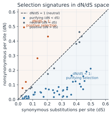
Synonymous vs. non-synonymous substitutions#
The genetic code is redundant: most amino acids are encoded by several codons, so a nucleotide change may or may not alter the protein.
A synonymous substitution changes the codon but not the amino acid it specifies (for example GGA to GGG, both glycine); because the protein is unchanged, it is approximately invisible to selection and evolves close to neutrally.
A non-synonymous substitution changes the encoded amino acid (for example GGA glycine to GAA glutamate); it alters the protein and is therefore exposed to selection, which can remove it or promote it.
The neutral background rate of synonymous change gives us a built-in control against which to measure the fate of non-synonymous change.
Defining , , and #
To make the comparison fair we normalize by opportunity, because a gene has more sites where a mutation would be non-synonymous than where it would be synonymous. Let be the number of synonymous substitutions per synonymous site and the number of non-synonymous substitutions per non-synonymous site. The quantity of interest is their ratio, Since estimates the neutral rate, measures how selection has scaled the rate of protein-changing substitutions relative to neutral expectation:
- — purifying (negative) selection: most amino-acid changes are deleterious and are removed, so is suppressed below .
- — neutral evolution: amino-acid changes fix at the same rate as synonymous ones.
- — positive (diversifying) selection: amino-acid changes are favored and fix faster than neutral, a classic signal at antigenic sites and drug-resistance loci in pathogens.
Purifying selection dominates most genes, so a gene-wide well below 1 is typical; values above 1 are the striking exception worth hunting for.
Codon models and where selection hides#
Simply counting differences (as below) ignores multiple hits at the same site and the structure of the genetic code, so modern analyses fit codon-substitution models by maximum likelihood, estimating as a model parameter along the branches of a phylogeny. Averaging over a whole gene is conservative: a handful of positively selected codons can be swamped by many conserved ones. Site models allow to vary across codons and test whether a subset of sites has , pinpointing, say, the antigenic residues of an influenza surface protein. Branch models let vary across lineages, testing whether selection intensified on a particular branch — for instance, the emergence of a drug-resistant clade. These tests compare a model that permits against one that does not, a nested-model comparison evaluated with the framework of hypothesis testing.
Worked example#
Suppose we align two homologous pathogen sequences of 300 codons (900 nucleotides) and, using the code’s structure, classify the sites and count observed differences:
- Synonymous sites: ; observed synonymous differences: .
- Non-synonymous sites: ; observed non-synonymous differences: .
The crude proportions of differences are Taking these proportions directly as rough distances gives An of about 0.5 points to purifying selection: protein-changing substitutions accumulate at half the neutral rate, so on average amino-acid changes here are mildly deleterious. A rigorous analysis would apply a multiple-hits correction (such as Nei–Gojobori’s Jukes–Cantor step) to convert and into and , but the sign of the signal, , is already clear.
In code#
The snippets reproduce the worked counts and the crude ratio, then point to library routines that add the proper corrections.
R#
# Crude omega from counts (matches the worked example)
Sd <- 18; S <- 225   # synonymous diffs and sites
Nd <- 27; N <- 675   # non-synonymous diffs and sites
pS <- Sd / S; pN <- Nd / N
omega <- pN / pS
omega                 # 0.5  -> purifying selection

# Proper estimation from an alignment with seqinr's Nei-Gojobori kaks():
# library(seqinr)
# aln <- read.alignment("genes.fasta", format = "fasta")
# kaks(aln)$ka / kaks(aln)$ks   # ka = dN, ks = dS
# (ape can read/handle trees; codon-model fitting lives in tools like codeml/PAML)
Python#
# Crude omega from counts
Sd, S = 18, 225   # synonymous diffs, sites
Nd, N = 27, 675   # non-synonymous diffs, sites
pS, pN = Sd / S, Nd / N
omega = pN / pS
print(round(omega, 3))   # 0.5 -> purifying selection

# Proper Nei-Gojobori estimation from an alignment with Biopython:
# from Bio import SeqIO
# from Bio.codonalign.codonseq import cal_dn_ds, CodonSeq
# s1 = CodonSeq(str(rec1.seq)); s2 = CodonSeq(str(rec2.seq))
# dN, dS = cal_dn_ds(s1, s2, method="NG86")
# print(dN / dS)
0.5
Julia#
# Crude omega from counts (manual)
Sd, S = 18, 225      # synonymous diffs, sites
Nd, N = 27, 675      # non-synonymous diffs, sites
pS, pN = Sd / S, Nd / N
omega = pN / pS
println(round(omega, digits = 3))   # 0.5 -> purifying selection

# Optional Jukes-Cantor correction before taking the ratio:
jc(p) = -3/4 * log(1 - 4/3 * p)     # multiple-hits correction
omega_corrected = jc(pN) / jc(pS)
println(round(omega_corrected, digits = 3))   # ~0.49, same conclusion
Why it matters#
converts a protein alignment into a quantitative test for natural selection, using the gene’s own synonymous sites as an internal neutral yardstick. For pathogens this is directly actionable: flags the antigenic sites that let a virus escape immunity and the codons where drug resistance is being selected, telling surveillance and vaccine design exactly which residues are under evolutionary pressure.